Everything to know about echocardiography
There are several medical imaging techniques to analyze the functioning and structural abnormalities of the heart. Echocardiography is one such test that helps doctors identify problems with the pumping organ. This non-invasive imaging technique is usually done at a hospital or medical lab. Heart rhythm disorders like bradycardia (low heart beat rate), tachycardia (elevated heart rate), and atrial fibrillation demand an echocardiography test to investigate the reason behind these conditions.
What is echocardiography?
Echocardiography is an ultrasound imaging of the heart. It is also known as cardiac echography, echo test, heart ultrasound, and heart sonogram. Echocardiography uses either standard ultrasound or Doppler ultrasound waves to produce live images of the heart. The picture thus formed is called an echocardiogram. An echocardiograph is considered a more accurate examination than the other heart tests in determining the structure and function of the organ.
What does an echocardiography help with?
Echocardiography can reveal a lot about the condition of the heart and help the physician diagnose several cardiac problems. A doctor may suggest this test if the patient complains of irregular heartbeat, chest pain, or shortness of breath.
Purpose of the test
Echocardiography is performed to examine the following:
The heart valve function
Any valve leakage
Heart valve narrowing
Size of the heart, any variation in the chamber size, dilation, or wall thickening.
Blood clots in the chambers or valves
Pericardial fluid accumulation
Blockage or narrowing of the aorta, which is the main artery that pumps blood from the heart to other parts of the body.
The contraction and relaxation of heart chambers during the pumping of blood
Heart pressure
Hear muscle damage
Types of echocardiography
The four types of echocardiography are detailed below:
Transthoracic echocardiogram or TTE
This is the most commonly performed type of echocardiography. The doctor will place a device called a transducer on the chest above the position of the heart. This sends ultrasound waves to the heart, and a computer connected to it produces an image of the heart using sound waves that bounce back. The person who undergoes the test may be given a contrast dye to get a better picture of the organ. Transthoracic echocardiography is usually performed to check the blood flow through the heart and valves.
Transesophageal echocardiogram or TEE
Transesophageal echocardiography is the best option when the normal heart ultrasound test does not provide any detailed information about the condition the doctor is looking for. In this procedure, the physician inserts a small transducer into the throat and is guided to reach near the heart through the esophagus. This will help to get a better image of the organ, especially the chambers which are not clear on the normal echocardiogram.
Stress echocardiogram
Here, transthoracic echocardiography is used to produce a stress echocardiogram. The images are taken before and after doing exercise at the clinic. This helps to check the response of the heart to physical activity or stress. Stress echocardiography is usually recommended if the doctor suspects coronary artery disease.
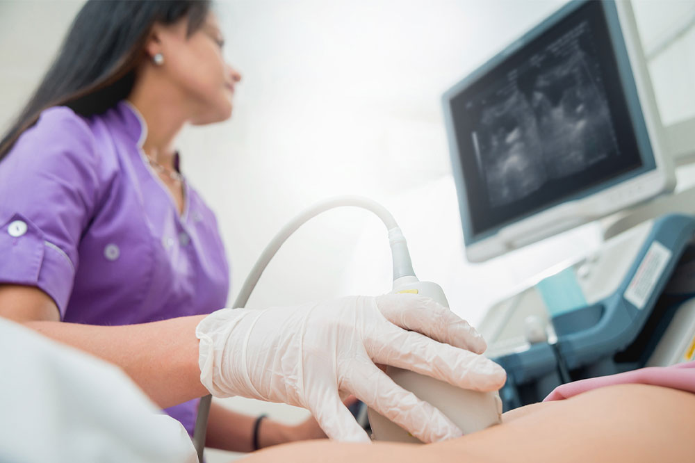
Fetal echocardiogram
Fetal echocardiography is done during pregnancy to check the fetus’s heart condition. The doctor moves an ultrasound wand over the pregnant woman’s belly during this test to observe the baby’s heart.
Techniques employed in echocardiography
Echocardiography employs the following techniques to create an image of the heart:
2D ultrasound
This is the most commonly used technique. It produces a 2D image of the heart, which appears as pieces on the computer. These are often combined to create a 3D image.
3D ultrasound
A 3D echocardiography helps to see the parts of the heart from different angles. It is often done to see a detailed image of the lower left chamber of the organ.
Doppler ultrasound
This is performed to check the rate and direction of blood flow. The sound waves transmitted to the heart change their pitch when they reflect off the blood cells’ surface. These changes, called Doppler signals, measure the speed and analyze the blood flow direction.
Color Doppler ultrasound
This technique is the same as the Doppler ultrasound but with the difference that this test uses a dye, which is inserted into the patient, to study the different directions of blood flow.
Strain imaging
The strain imaging technique is used to monitor the heart muscle movement.
Color flow imaging
In this method, a contrast dye is administered to the patient and then tracked during the imaging procedure. This substance helps identify leaks in the heart valves.
Risks
Echocardiography is a safe and non-invasive procedure that uses ultrasound, which means there is no risk to the body. However, a few people may experience an allergic reaction to the contrast dye used in color Doppler ultrasound.
Conclusion
Echocardiography is the most reliable imaging test to assess heart health. It provides information on a wide variety of heart disorders. The cost of the test varies depending on the type, technique used, and the hospital.